
Patients should be warned of this.read more read lessĪbstract: Syndesmosis injuries occur when there is a disruption of the distal attachment of the tibia and fibula.
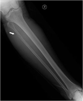
MAISONNEUVE FRACTURE SERIES
This series shows that there is a significant incidence of soft-tissue complications with the use of tightrope fixation and subsequent need for removal. Functional outcome was good and patients were satisfied. In all cases the syndesmosis was reduced and held, even after device removal. Average time to weightbearing was six weeks (range 4-8). Histological examination revealed refractile material within giant cells, suggestive of foreign-body reaction. In two cases the device caused soft-tissue irritation with granuloma formation, necessitating subsequent removal, one after six months, and the other after ten months. Fractures were treated according to AO-ASIF principles and the tightrope applied through a plate in three cases and directly through the fibula in three cases. These included four Weber C fractures, one Maisonneuve fracture and one isolated diastasis without fracture. Six patients were treated for ankle diastasis using the tightrope. We are the first to report complications with the use of this device. Early clinical results indicate that it can remain in situ indefinitely without complications. It would seem reasonable to place a syndesmotic screw at 2.0 cm above tibiotalar joint.read more read lessĪbstract: Fixation of ankle syndesmosis injury with a fibre-wire tightrope has previously been reported. Two-tailed t-test comparing screw fixation at 2.0 cm and 3.5 cm indicated less syndesmotic widening with screw placed at 2.0 cm (P = 0.07). Two-tailed t-test comparing no fixation with fixation at 2.0 cm indicated less syndesmotic widening with screw placed at 2.0 cm (P = 0.04). Torsional force, the degree of rotation and the amount of syndesmotic widening were quantitated. The proximal tibia was internally rotated while the ankle was held fixed until syndesmotic, bony, or hardware failure occurred. The ankle was placed in neutral with 15 degrees of pronation and a load of 150 pounds and a strain gauge anchored medially and laterally. After ligament section and syndesmosis fixation, each leg was then jig mounted with transfixing wires through the proximal tibia and calcaneus. In group II (7 pairs), the syndesmosis in each left leg was fixed at 3.5 cm above the tibiotalar joint and the right leg syndesmosis was fixed at 2.0 cm above the tibiotalar joint. In group I (10 pairs), the left legs were tested without any syndesmotic fixation and the right legs were tested with the syndesmosis fixed at 2.0 cm above the tibiotalar joint. The paired cadaver legs were tested in two groups. The legs were then dissected to expose the syndesmosis/interosseous membrane. Legs of 17 embalmed cadavers underwent knee disarticulation. The aim/purpose of this study was to demonstrate the optimal level of syndesmotic screw placement before creation of a Maisonneuve fracture. We found that the ruptured deltoid ligament did not need to be repaired if the lateral side was anatomically and rigidly fixed in the Maisonneuve fracture, restoration of the fibular length was as important as stabilization of the fracture with the use of the suprasyndesmotic screw, walking was permissible with the screw in situ conforming the plate to the bend of the lateral malleolus was essential and as much as two millimeters of lateral residual displacement of the lateral and medial malleoli was compatible with a satisfactory result, as was a similar displacement of the talus provided there was anatomical restoration of the lateral side.read more read lessĪbstract: At the present time, syndesmotic screw fixation is recommended when there is a tibiofibular diastasis, a Maisonneuve fracture, or syndesmotic instability after fixation of distal tibia-fibula fractures. Less satisfactory results were noted in the more severely injured ankles. After an average follow-up of three and one-half years, the results were satisfactory in 90 per cent. Abstract: One hundred and fifty patients with a displaced fracture of the ankle caused by external rotation-abduction forces were treated by open reduction and rigid internal fixation.


 0 kommentar(er)
